


New Pathway to Treating Adult Amblyopia
 Researchers at the Massachusetts Institute of Technology have uncovered a promising new approach to treating amblyopia in adults, challenging the long-held belief that the condition can only be effectively treated during early childhood.
The study, published in Cell Reports and led by former graduate student Madison Echavarri-Leet under Professor Mark Bear at MIT's Picower Institute for Learning and Memory, demonstrates that temporarily anesthetising the retina of the amblyopic eye for just two days can restore the brain's visual response to that eye, even in adulthood.
Breaking the Critical Period Barrier
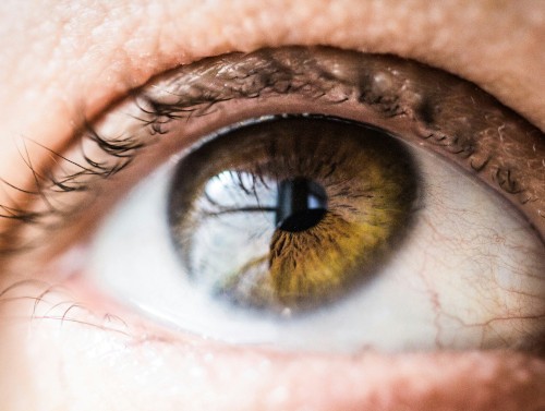
Amblyopia, commonly known as lazy eye, affects approximately 3% of the global population. Current interventions have only been effective during infancy and early childhood while neural connections are still being formed. The condition occurs when impaired vision in one eye during development causes the brain's visual system to favour the other eye, leaving the amblyopic eye less capable even after the original impairment is corrected.
The MIT research offers hope for the estimated millions of adults living with amblyopia who have been considered beyond treatment.
The Mechanism: Burst Mode Firing
The breakthrough lies in understanding how retinal inactivation triggers visual recovery. The research team discovered that blocking inputs from a retina to neurons in the lateral geniculate nucleus (LGN) causes those neurons to fire synchronous "bursts" of electrical signals to downstream neurons in the visual cortex.
These burst patterns are similar to neural activity that occurs in the visual system before birth, which guides early synaptic development. The researchers found that this bursting depends on T-type calcium channels in the LGN neurons.
To confirm the importance of this mechanism, the team genetically knocked out these calcium channels in mice. When the channels were eliminated and bursting was disrupted, anesthetising the non-amblyopic eye could no longer help amblyopic mice, proving that bursting is necessary for the treatment to work.
A Significant Clinical Advantage
Perhaps most importantly for clinical application, the researchers discovered that the treatment works when applied directly to the amblyopic eye itself, not just the fellow eye.
In experiments using tetrodotoxin (TTX) to temporarily inactivate the retina, the team found that after a week, the ratio of input from each eye was much more even in mice that received treatment compared to those left untreated, indicating that the amblyopic eye's input in the brain rose to be at parity with input from the non-amblyopic eye.
"If it does [work in humans], it's a pretty substantial step forward because it would be reassuring to know that vision in the good eye would not have to be interrupted by treatment," Professor Bear noted. "The amblyopic eye, which is not doing much, could be inactivated and 'brought back to life' instead".
Building on Previous Research
This study builds on the lab's 2021 research, which showed that anesthetising the non-amblyopic eye could improve vision in the amblyopic one – an approach analogous to the patching treatment used in childhood. Those 2021 findings have now been replicated in adults of multiple species.
Earlier work from 2016 demonstrated that temporarily anesthetising both retinas could restore vision loss in amblyopia, establishing proof of concept for the broader approach.
Safety Considerations
Research on retinal safety has shown encouraging results. Studies examining retinal and optic nerve integrity following monocular inactivation found no evidence of gross histopathological abnormalities following inactivation for 10 days, nor evidence of degeneration of axons or loss of myelin in the optic nerve.
Next Steps and Clinical Implications
The researchers emphasise that significant work remains before this approach can be considered for human trials. Professor Bear stressed that "especially with any invasive treatment, it's extremely important to confirm the results in higher species with visual systems closer to our own".
Recent work in rhesus monkeys has shown promise, with low doses of tetrodotoxin promoting recovery in the amblyopic eye without compromising the function or structure of the treated eye.
The research team, which also includes Tushar Chauhan, Teresa Cramer, and Ming-fai Fong, wrote that they are "cautiously optimistic that these findings may lead to a new treatment approach for human amblyopia, particularly given the discovery that silencing the amblyopic eye is effective".
Implications for Practice
For eyecare professionals, this research represents a significant shift in understanding amblyopia's treatment potential. While clinical applications are still years away, the findings suggest that adult patients with amblyopia may not be beyond help, particularly those with deprivation amblyopia – typically the most severe and treatment-resistant form of the condition.